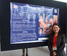
Dermatofibromas- conventional versus polarized dermoscopy
 I was fortunate to be chosen to present at the Free Communications Session at the Congress of the International Dermoscopy Society (Naples, Italy). I presented on behalf of co-investigators Drs. S. Taliercio, C. Salaro, S. Dusza, P. Chu and A. Marghoob on our paper on the "Conventional and polarized dermoscopy features of dermatofibroma". Basically, what we found from the study was that there are differences with the resulting image when using conventional versus polarized dermoscopy. These differences may become significant in that while many dermoscopy courses and atlases feature photos of lesions taken with conventional dermoscopy, many neophyte dermoscopists are starting out with polarized dermoscopes.
This is the abstract of the talk, which has since also been published in the Archives of Dermatology (see Publications link):
OBJECTIVE: To evaluate dermoscopic features and patterns of dermatofibromas using conventional and polarized light dermoscopy.
DESIGN: Dermatofibromas were imaged using conventional nonpolarized contact dermoscopy (NPD), polarized contact dermoscopy (PCD), and polarized noncontact dermoscopy, followed by evaluation and comparison of dermoscopic features of the lesions.
SETTING: Dermatology clinic specializing in pigmented lesions. Patients Fifty patients with dermatofibromas.
RESULTS: The most common features of dermatofibromas observed with NPD and PCD were central white scarlike patches (37 [74%] and 42 [84%], respectively), brown globulelike structures (21 [42%] and 22 [44%]), vascular structures (24 [48%] and 22 [44%]), and a peripheral fine pigmented network (36 [72%] for both). A newly described feature observed with PCD was a central white patch characterized by shiny white streaks. With polarized noncontact dermoscopy, the most characteristic feature was a central pink hue or "vascular blush" (44 [88%]) and visibility of blood vessels (41 [82%]). The most common pattern identified with NPD and PCD was the combination of a peripheral pigmented network and a central white patch in 28 (56%) and 31 (62%) of lesions, respectively. With polarized noncontact dermoscopy, the most common pattern was a central pink hue with a peripheral pigmented network (23 [46%]). There was good to excellent agreement when comparing NPD with PCD images, but there was a variable level of agreement when polarized noncontact dermoscopy images were compared with NPD and PCD images.
CONCLUSIONS: Conventional and polarized light dermoscopy are not equivalent but may be complementary. This study highlights some salient differences. We were able to identify new dermoscopic features and patterns not previously described with conventional dermoscopy. These new criteria can aid in the diagnosis of dermatofibroma. (At the meeting with Dr. Anne Amon and Dr. Vangie Handog, dermatologists from Manila)


No comments:
Post a Comment