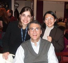
This was my first time to participate as a faculty at a two hour forum at the American Academy of Dermatology Annual Meeting (San Franciscio, 2006). They spelled my name wrong however in the program (!!!)
My thanks to my two mentors, Dr. Allan C. Halpern (chief of the Dermatology Service at Memorial Sloan-Kettering Cancer Center, NY, USA) and Dr. Patricia L. Myskowski (also a consultant at MSKCC). They understood how nervous I was for this session, and gave me lots of support! It was very well attended as many dermatologists are dealing more and more with patients suffering from these adverse events as the use of anti-EGFR cancer treatment becomes more widespread.
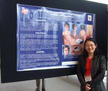
This is the abstract for this session. We have also published on this topic at the Journal of the American Academy of Dermatology (see Publications link).
SKIN EFFECTS OF BIOLOGICALLY TARGETED CANCER THERAPIES
Anna Liza C Agero MD, Allan C. Halpern MD, Patricia Myskowski MD
Memorial Sloan-Kettering Cancer Center, New York, NY, USA
SHORT ABSTRACT
One of the recent exciting advances in medicine has been the development of novel biologically targeted agents that interfere with specific molecular pathways affecting the evolution of cancer. Some examples of these new class of anti-neoplastic agents include monoclonal antibodies and inhibitors of intracellular signaling pathways (receptor and tyrosine kinase inhibitors). A number of unique and dramatic dermatologic side-effects have been associated with the use of these biotherapeutic agents. This forum will summarize the spectrum of cutaneous reactions to some of these agents, focusing on the epidermal growth factor receptor (EGFR) inhibitors and some multi-targeted kinase inhibitors, based primarily on our experience at Memorial Sloan-Kettering Cancer Center supplemented with reports from literature. This forum will identify these dermatologic adverse reactions, describe their clinical and histopathologic features, as well as possible mechanisms and some suggested treatments.
LONG ABSTRACT
One of the recent exciting advances in medicine has been the development of novel biologically targeted agents that interfere with specific molecular pathways affecting the evolution of cancer. Some examples of these new class of anti-neoplastic agents include monoclonal antibodies and inhibitors of intracellular signaling pathways (receptor and tyrosine kinase inhibitors). This forum summarizes the spectrum of cutaneous reactions to some of these agents, focusing on the epidermal growth factor receptor (EGFR) inhibitors and some multi-targeted kinase inhibitors, based primarily on our experience at Memorial Sloan-Kettering Cancer Center supplemented with reports from literature.
Epidermal growth factor receptor (EGFR) inhibitors are members of this new class of anti-neoplastic agents. These agents were developed for therapeutic blockade of the epidermal growth factor receptor function, which has been shown to play a significant role in signal transduction pathways affecting cancer growth and spread. These targeted therapies have the potential of offering efficacy without the major toxicities associated with conventional chemotherapeutic agents. The EGFR-targeted antibody Cetuximab, and the small tyrosine kinase inhibitors Erlotinib and Gefitinib have recently received approval from the FDA for treatment of patients with colorectal and non-small cell lung cancer refractory or intolerant to chemotherapy.1,2 In particular, Erlotinib therapy has been shown to improve survival in patients with advanced or refractory non-small cell lung cancer compared to placebo.3 Treatment with Gefitinib has also demonstrated clinically meaningful improvements in disease-related symptoms and health related quality of life in patients with advanced non-small cell lung cancer. Erlotinib treatment is another anti-EGFR agent that has also shown improvement in patients’ health-related quality of life.4 Anti-EGFR therapies are currently being tested in other disease settings, and as their use become more prevalent, their side-effects profiles become clinically significant.
Anti-EGFR therapy is commonly associated with unique and dramatic dermatologic side-effects that appear to be secondary to inhibition of specific roles EGFR plays in skin biology. The most commonly encountered toxicity in patients is a characteristic skin rash that occurs in up to 85% of patients, while nail, hair and mucosal changes appear less frequently.5 The skin rash is characterized by sterile suppurative follicular lesions6,7, and is commonly described as “acne-like” and “acneiform.”3 The face, upper chest and back are the most frequently affected areas.5 The follicular rash often commences between days ten and fourteen of therapy,7,8 and is maximal by weeks three to five.9 A dose-related reaction is also seen, with higher incidence and more severe rash noted at higher dose levels.7,10 The skin reaction is generally mild and well tolerated, usually accompanied only by minimal discomfort or pruritus,8,11-13 but may rarely be more severe and widespread.14 The rash is rarely dose-limiting, and in a number of studies, skin toxicity did not exceed Grade 2 in severity.15-17 However, there are cases wherein a patient was severely distressed by the cosmetic effects, and chose to withdraw from treatment although the rash was asymptomatic.14
Currently, there is no specific evidence-based treatment for the pustular-follicular rash caused by the anti-EGFR agents. Patients who develop mild to moderate skin toxicity are recommended to continue treatment without dose modification. When intervention is indicated, patients have been treated empirically with drying agents, retinoids, steroids and antibiotics.8 However, attempts at clinical prevention or improvement of the rash have not shown consistent benefit.6,18,19
Histopathology studies of the pustular-follicular lesions have revealed two major patterns of reactions: a moderate superficial dermal inflammatory cell infiltrate surrounding hyperkeratotic and ectatic follicular infundibula, particularly in the upper portion of the hair follicle, and a superficial neutrophilic suppurative folliculitis with associated rupture of epithelial lining.6,18,20 The exact mechanism for the ‘acne-like’ and folliculo-centricity of the rash is still not clear, though it is agreed that it is a consequence of inhibition of EGFR signaling on epidermal and adnexal epithelium. Several pre-clinical studies have shown an essential role for EGFR activation in keratinocyte proliferation with studies of EGFR-knockout mice showing skin and hair abnormalities.21
Significantly, occurrence of the rash has been linked to better clinical outcomes,22 such that studies investigating dose escalation of Erlotinib until occurrence of Grade 2 or maximally tolerated rash are underway in order to determine clinical benefit at the maximally tolerated dose.23,24 However, more data are needed to confirm a possible relationship between rash (and severity of rash) with clinical response.
Other changes associated with anti-EGFR therapy include painful nail paronychia characterized as a periungual granulation-type of paronychia 25 or friable pyogenic granuloma-like changes18, coupled with erythema, tenderness, swelling and fissuring of lateral nail folds and/or distal finger tufts.18,26,27 Paronychia often develops after 2-4 months of treatment,27 but with a much lower incidence (12%) than rash.26 Changes with hair growth have also been noted, with reports of curlier, finer and more brittle hair on the scalp and extremities, and slowed growth of beard hair.28,29 Extensive growth of both eyelashes and eyebrows have also been reported.30 Other dermatologic changes observed are skin dryness that develops after onset of rash, mild to moderate mucositis, and hypersensitivity reactions.5
Other biologically targeted therapies include the multi-targeted kinase inhibitors: Imatinib (inhibitor of platelet-derived growth factor (PDGF), stem cell factor (SCF), c-kit, and Bcr-Abl), BAY 43-9006/Sorafenib (inhibitor of Raf/MEK/ERK pathway, VEGFR-2 &3 , FLT-3, p38, PDGFR and c-kit) and SU11248/ Sunitinib (inhibitor of VEGFR-2, PDGFR, FLT3 TKr and c-kit).5 Imatinib is FDA approved for refractory chronic myeloid leukemia, BAY 43-9006 for advanced renal cell carcinoma, and SU11248 is undergoing clinical trials as a treatment for gastrointestinal stromal tumor and metastatic renal cell carcinoma (www.fda.gov).
Treatment with Imatinib has been associated with a wide range of skin reactions, with 30-40% incidence of non-specific self-limited skin rash, and rare reports of more severe drug reactions such as vasculitis, Stevens-Johnson Syndrome, toxic epidermal necrolysis and acute generalized exanthematous pustulosis.5
Changes in skin and hair pigmentation have been observed during treatment with Imatinib and SU11248, with reports of dose-dependent and reversible local and diffused skin hypo- or depigmentation and variable reports of hair depigmentation and/or repigmentation.5 Pigmentation changes may be attributed to selectivity to the C-kit receptor which plays a regulatory role in melanocyte development.
Subungual splinter hemorrhages have been a common side effect (30-60% of patients) of patients taking SU11248 and BAY 439006. These have not been associated with thrombotic and embolic events, and may be secondary to inhibition of VEGFR.5
As use of these targeted agents become more prevalent, it is important that dermatologists be able to properly diagnose these side-effects, and differentiate them from other skin disorders. These dermatologic reactions may be relatively common and generally mild, but may also cause clinical distress to the patient.
REFERENCES
1. Kim ES, Khuri FR, Herbst RS. Epidermal growth factor receptor biology (IMC-C225). Curr Opin Oncol. 2001;13:506-513.
2. De Bono JS, Rowinsky EK. The ErbB receptor family: a therapeutic target for cancer. Trends Mol Med. 2002;8:S19-26.
3. Perez-Soler R, Delord JP, Halpern A, et al. HER1/EGFR inhibitor-associated rash: future directions for management and investigation outcomes from the HER1/EGFR inhibitor rash management forum. Oncologist. 2005;10:345-356.
4. Fallowfield LJ, Harper P. Health-related quality of life in patients undergoing drug therapy for advanced non-small-cell lung cancer. Lung Cancer. 2005;48:365-377.
5. Robert C, Soria JC, Spatz A, et al. Cutaneous side-effects of kinase inhibitors and blocking antibodies. Lancet Oncol. 2005;6:491-500.
6. Cohen R, Falcey J, Paulter V, Fetzer K, Waksal H. Safety profile of the monoclonal antibody (MoAb) IMC-C225, an anti-epidermal growth factor receptor (EGFr) used in the treatment of EGFr-positive tumors. Proc Am Soc Clin Oncol. 2000;19:474a Abs. 1862.
7. Ranson M, Hammond LA, Ferry D, et al. ZD1839, a selective oral epidermal growth factor receptor-tyrosine kinase inhibitor, is well tolerated and active in patients with solid, malignant tumors: results of a phase I trial. J Clin Oncol. 2002;20:2240-2250.
8. Perez-Soler R, Chachoua A, Hammond LA, et al. Determinants of tumor response and survival with erlotinib in patients with non--small-cell lung cancer. J Clin Oncol. 2004;22:3238-3247.
9. Rowinsky EK, Schwartz GH, Gollob JA, et al. Safety, pharmacokinetics, and activity of ABX-EGF, a fully human anti-epidermal growth factor receptor monoclonal antibody in patients with metastatic renal cell cancer. J Clin Oncol. 2004;22:3003-3015.
10. Herbst RS, Maddox AM, Rothenberg ML, et al. Selective oral epidermal growth factor receptor tyrosine kinase inhibitor ZD1839 is generally well-tolerated and has activity in non-small-cell lung cancer and other solid tumors: results of a phase I trial. J Clin Oncol. 2002;20:3815-3825.
11. Walon L, Gilbeau C, Lachapelle JM. Acneiform eruptions induced by cetuximab. Ann Dermatol Venereol. 2003;130:443-446.
12. Schwartz G, Dutcher JP, Vogelzang NJ, et al. Phase 2 clinical trial evaluating the safety and effectiveness of ABX-EGF in renal cell cancer (RCC). Proc Am Soc Clin Oncol. 2002;21:24 Abs. 91.
13. Albanell J, Rojo F, Averbuch S, et al. Pharmacodynamic studies of the epidermal growth factor receptor inhibitor ZD1839 in skin from cancer patients: histopathologic and molecular consequences of receptor inhibition. J Clin Oncol. 2002;20:110-124.
14. Yamamoto DS, Viale PH, Zhao G. Severe acneiform rash. Clin J Oncol Nurs. 2004;8:654-656.
15. Kollmannsberger C, Schittenhelm M, Honecker F, et al. Epidermal growth factor receptor (EGFR) antibody EMD 72000 in combination with paclitaxel (P) in patients (pts) with EGFR-positive advanced non-small cell lung cancer (NSCLC): A phase-I study. Proc Am Soc Clin Oncol. 2003;22:627 Abs. 25206.
16. Trarbach T, Beyer T, Schleucher N, et al. A randomized phase I study of the humanized anti-epidermal growth factor receptor (EGFR) monoclonal antibody EMD 72000 in subjects with advanced gastrointestinal cancers. Journal of Clinical Oncology, 2004 ASCO Annual Meeting Proceedings (Post-Meeting Edition). 2004;22:3018.
17. Herbst RS, LoRusso PM, Purdom M, Ward D. Dermatologic side effects associated with gefitinib therapy: clinical experience and management. Clin Lung Cancer. 2003;4:366-369.
18. Busam KJ, Capodieci P, Motzer R, Kiehn T, Phelan D, Halpern AC. Cutaneous side-effects in cancer patients treated with the antiepidermal growth factor receptor antibody C225. Br J Dermatol. 2001;144:1169-1176.
19. Hidalgo M, Siu LL, Nemunaitis J, et al. Phase I and pharmacologic study of OSI-774, an epidermal growth factor receptor tyrosine kinase inhibitor, in patients with advanced solid malignancies. J Clin Oncol. 2001;19:3267-3279.
20. Jacot W, Bessis D, Jorda E, et al. Acneiform eruption induced by epidermal growth factor receptor inhibitors in patients with solid tumours. Br J Dermatol. 2004;151:238-241.
21. Jost M, Kari C, Rodeck U. The EGF receptor - an essential regulator of multiple epidermal functions. Eur J Dermatol. 2000;10:505-510.
22. Saltz LB, Kies MS, Abbruzzese JL, Azarnia N, Needle M. The presence and intensity of the cetuximab-induced acne-like rash predicts increased survival in studies across multiple malignancies. Proc Am Soc Clin Oncol. 2003;22:204 Abs. 817.
23. Brewer CJ, Suh JH, Stevens GHJ, et al. Phase II trial of erlotinib with temozolomide and concurrent radiation therapy in patients with newly-diagnosed glioblastoma multiforme. Journal of Clinical Oncology, 2005 ASCO Annual Meeting Proceedings. 2005;23:16S Abs.1567.
24. Mita CA, Schwartz G, Mita MM, et al. A pilot, pharmacokinetic (PK), and pharmacodynamic (PD) study to determine the feasibility of intrapatient dose escalation to tolerable rash and the activity of maximal doses of erlotinib (E) in previously treated patients with advanced non-small cell lung cancer (NSCLC). Journal of Clinical Oncology, 2005 ASCO Annual Meeting Proceedings. 2005;23.
25. Boucher KW, Davidson K, Mirakhur B, Goldberg J, Heymann WR. Paronychia induced by cetuximab, an antiepidermal growth factor receptor antibody. J Am Acad Dermatol. 2002;47:632-633.
26. Saltz LB, Meropol NJ, Loehrer PJ, Sr., Needle MN, Kopit J, Mayer RJ. Phase II trial of cetuximab in patients with refractory colorectal cancer that expresses the epidermal growth factor receptor. J Clin Oncol. 2004;22:1201-1208.
27. Monti M, Mancini LL, Ferrari B, Rahal D, Santoro A. Complications of therapy and a diagnostic dilemma case. Case 2. Cutaneous toxicity induced by cetuximab. J Clin Oncol. 2003;21:4651-4653.
28. Van Doorn R, Kirtschig G, Scheffer E, Stoof TJ, Giaccone G. Follicular and epidermal alterations in patients treated with ZD1839 (Iressa), an inhibitor of the epidermal growth factor receptor. Br J Dermatol. 2002;147:598-601.
29. Cohen EE, Rosen F, Stadler WM, et al. Phase II trial of ZD1839 in recurrent or metastatic squamous cell carcinoma of the head and neck. J Clin Oncol. 2003;21:1980-1987.
30. Dueland S, Sauer T, Lund-Johansen F, Ostenstad B, Tveit KM. Epidermal growth factor receptor inhibition induces trichomegaly. Acta Oncol. 2003;42:345-346.