Most Recent Posts

Saturday, August 4, 2007

Not dermatology-related, but just wanted to share this...


This amazing picture was featured at The Philippine Daily Inquirer (Sat, Aug 04, 2007)---

"BEAUTY AFTER RUIN. More than 16 years after its destructive eruption in 1991, Mount Pinatubo now offers this breathtaking crater-lake at its summit. INQUIRER/TONETTE T. OREJAS"

Are there differences between polarized light dermoscopy and immersion contact dermoscopy?

I also presented on the "Differences between polarized light dermoscopy and immersion contact dermoscopy for the evaluation of skin lesions" at the Free Communications Session at the Congress of the International Dermoscopy Society (Naples, Italy). I presented on behalf of co-investigators Drs. Benvenuto-Andrade C, Dusza SW, Agero AL, Scope A, Rajadhyaksha M, Halpern AC and Marghoob AA. This paper has since been published at the Archives of Dermatology (see Publications link).

I attach below the abstract:

OBJECTIVE: To evaluate dermoscopic features and patterns of skin lesions by using
conventional and polarized light dermoscopy (PD).

DESIGN: Observational study.

SETTING: Dermatology clinic at Memorial Sloan-Kettering Cancer Center.

PATIENTS: Ninety patients with skin lesions.

INTERVENTIONS: Skin lesions were imaged via conventional nonpolarized light contact dermoscopy (NPD), polarized light contact dermoscopy (PCD), and polarized light noncontact dermoscopy (PNCD).

MAIN OUTCOME MEASURES: The images from the 3 modalities were evaluated by 3 dermoscopists for colors, structures, and patterns. Level of agreement between modalities was assessed by percentage agreement and the kappa statistic. Qualitative differences between modalities were also assessed.

RESULTS: Ninety lesions comprising 55 melanocytic and 35 nonmelanocytic lesions were reviewed. There was excellent agreement for overall dermoscopic patterns between modalities, with kappa values ranging from 0.88 to 1.00. There was moderate to excellent agreement for most dermoscopic colors, with the exception of blue-white veil and pink (red) color. Most dermoscopic structures had fair to perfect agreement, with the exception of milialike cysts. Qualitative assessment suggested that melanin appeared darker and blue nevi had more shades of blue on PD compared with NPD images; vessels and red areas were better visualized with PD, suggesting that PD may be helpful in identifying malignancies; milialike cysts and comedolike openings were better visualized with NPD, suggesting that NPD is more helpful for identification of seborrheic keratosis; peppering, lighter colors, and blue-white areas were more evident under NPD, facilitating recognition of regression areas; and shiny-white streaks, possibly representing fibrosis, were seen more clearly under PD.

CONCLUSIONS: The capabilities of NPD, PCD, and PNCD are not equivalent, but complementary. Further studies are needed to evaluate the effect of these differences on clinical diagnosis.

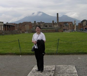
(Picture taken at the ruins of Pompeii, with Vesuvius in the background)

Saturday, July 28, 2007

Dermatofibromas- conventional versus polarized dermoscopy

I was fortunate to be chosen to present at the Free Communications Session at the Congress of the International Dermoscopy Society (Naples, Italy). I presented on behalf of co-investigators Drs. S. Taliercio, C. Salaro, S. Dusza, P. Chu and A. Marghoob on our paper on the "Conventional and polarized dermoscopy features of dermatofibroma". Basically, what we found from the study was that there are differences with the resulting image when using conventional versus polarized dermoscopy. These differences may become significant in that while many dermoscopy courses and atlases feature photos of lesions taken with conventional dermoscopy, many neophyte dermoscopists are starting out with polarized dermoscopes.

This is the abstract of the talk, which has since also been published in the Archives of Dermatology (see Publications link):

OBJECTIVE: To evaluate dermoscopic features and patterns of dermatofibromas using conventional and polarized light dermoscopy.

DESIGN: Dermatofibromas were imaged using conventional nonpolarized contact dermoscopy (NPD), polarized contact dermoscopy (PCD), and polarized noncontact dermoscopy, followed by evaluation and comparison of dermoscopic features of the lesions.

SETTING: Dermatology clinic specializing in pigmented lesions. Patients Fifty patients with dermatofibromas.

RESULTS: The most common features of dermatofibromas observed with NPD and PCD were central white scarlike patches (37 [74%] and 42 [84%], respectively), brown globulelike structures (21 [42%] and 22 [44%]), vascular structures (24 [48%] and 22 [44%]), and a peripheral fine pigmented network (36 [72%] for both). A newly described feature observed with PCD was a central white patch characterized by shiny white streaks. With polarized noncontact dermoscopy, the most characteristic feature was a central pink hue or "vascular blush" (44 [88%]) and visibility of blood vessels (41 [82%]). The most common pattern identified with NPD and PCD was the combination of a peripheral pigmented network and a central white patch in 28 (56%) and 31 (62%) of lesions, respectively. With polarized noncontact dermoscopy, the most common pattern was a central pink hue with a peripheral pigmented network (23 [46%]). There was good to excellent agreement when comparing NPD with PCD images, but there was a variable level of agreement when polarized noncontact dermoscopy images were compared with NPD and PCD images.

CONCLUSIONS: Conventional and polarized light dermoscopy are not equivalent but may be complementary. This study highlights some salient differences. We were able to identify new dermoscopic features and patterns not previously described with conventional dermoscopy. These new criteria can aid in the diagnosis of dermatofibroma.

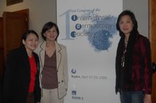
(At the meeting with Dr. Anne Amon and Dr. Vangie Handog, dermatologists from Manila)

Sunday, July 22, 2007

A stand alone autologous cell harvesting device - treatment for Vitiligo?


ReCell® is a novel single-use medical device developed as an 'off the shelf' kit for harvesting autologous skin cells taken from a thin split thickness biopsy. The process provides an immediate cell population composed of keratinocytes, melanocytes, fibroblasts and Langerhans cells harvested from the epidermal-dermal junction for delivery onto the wound surface in order to promote effective wound healing. ReCell® has been used to treat smaller wounds such as small burns and scalds, donor sites, glabrous injuries, congenital nevi, hypopigmentation, chronic wounds and prophylactically in cosmetic rejuvenation procedures. The procedure is simple. Once processed, the cell suspension is available for immediate use and can cover a wound many times the area of the donor biopsy. As the ReCell® device enables cell processing at the site of treatment without the use of specialised laboratory staff, the process is both cost and time efficient.

Our interest in ReCell® stems from its ability to enable melanocyte repopulation which may result in the reintroduction of pigmentation into hypopigmented skin such as in hypopigmented scars, iatrogenic hypopigmentation and Vitiligo. At St George Hospital (NSW, Australia), we are currently conducting a randomized double blind study comparing ReCell® versus minigrafting for patients with stable vitiligo who have previously failed medical treatment. My co-investigators include Dr. John Le Guay and Dr. Richard Wittal of the Skin & Cancer Foundation and A/Prof Dedee Murrell and Dr. Linda Martin (St. George Hospital). We have currently recruited 14 out of a target of 24 patients, and the study is still open for interested patients. The entire procedure is performed by just myself and our trial nurse (Lesley Rhodes SRN), as the whole process does not require specialized laboratory staff. Patients also find it convenient as the on-site processing allows immediate application.


Sunday, July 15, 2007

Minocycline for EGFR-inhibitor associated skin rash


I was fortunate to be chosen as one of 20 presenters at the Residents & Fellows Symposium at the Annual Meeting of the American Academy of Dermatology (Washington DC, 2007). My presentation was on "PROPHYLAXIS WITH SYSTEMIC MINOCYCLINE AND TOPICAL TAZAROTENE (A RETINOID) FOR THE CETUXIMAB ASSOCIATED ACNE-LIKE ERUPTION". I presented on behalf of co-investigators Drs. Alon Scope, Stephen W. Dusza, Patricia L. Myskowski, Jocelyn Lieb, Leonard Saltz, Nancy E. Kemeny and Allan C. Halpern.

Cetuximab, an epidermal growth factor receptor inhibitor, is a biologically targeted agent that has been approved by the FDA for treatment of chemotherapy resistant/intolerant patients with metastatic colorectal carcinoma and unresectable squamous cell cancer of the head and neck. Cetuximab is associated with a characteristic dose-related follicular-pustular (acne-like) eruption affecting up to 90% of patients that is severe or dose-limiting in 10-20% of patients. Presently, there are no specific evidence-based treatments available for the inflammatory eruption. Patients are typically treated with dose modification, and empirically with drying agents, retinoids, steroids and antibiotics, but without evidence of consistent benefit.

This double-blinded randomized placebo-controlled single-center study aimed to assess the utility of topical tazarotene, oral minocycline or combination therapy for prevention of cetuximab-related rash. These agents were chosen based on anecdotal reports of their efficacy in cetuximab-related rash and their potential impact on follicular integrity and inflammation.

Patients with a diagnosis of colorectal cancer initiating cetuximab therapy were randomized to receive half-face twice a day topical therapy with tazarotene cream 0.01% and either oral minocycline (100mg daily) or oral placebo for two months. The primary endpoint in the study was the difference in total face lesion counts between minocycline- and placebo-treated patients at week 8. Secondary endpoints included the difference in total face lesion counts between minocycline- and placebo-treated patients at weeks 1-4 and difference in lesion counts between tazarotene-treated and observation sides of the face at weeks 1-8.

Forty eight eligible patients were randomly assigned to minocycline (n=24) or placebo (n=24). Median age was 44 years (range 38-83), with M: F (2:1) and 84% were white non-Hispanic. There was no significant difference between total facial lesions counts at week 8 between these groups. However, lesion counts were significantly attenuated in patients on minocycline at weeks 1-4. In 4 patients in the placebo- and in none in the minocycline-arm, cetuximab treatment was interrupted due to grade 3 skin adverse events. There was limited clinical benefit to tazarotene application.

We concluded that prophylaxis with minocycline was effective in decreasing severity of the acneiform rash during the first weeks of cetuximab therapy, suggesting that short-term minocycline prophylaxis for patients starting on cetuximab is beneficial.

This study was supported by an educational grant from Bristol-Myers Squibb Company, US, and we have submitted our findings for publication.

(Reunion of the MSKCC team at the AAD meeting in Washington DC. Top row, L-R, Ms Daphne Demas, Dr. Philip Spencer, Dr. Cristiane Benvenuto-Andarade, me, Dr. Patricia Myskowski, Dr. Alon Scope, Steve Dusza, Bottom row, L-R, Jules Lipoff, Dr. Jason Chen, Dr. Allan Halpern, Dr. Jocelyn Lieb)

Friday, July 13, 2007

Skin cancer biopsy without a knife


On Nov. 2 2006, "Today" host Matt Lauer spoke with Dr. Allan Halpern, chief of dermatology at Memorial Sloan-Kettering Cancer Center (MSKCC), about a new medical tool, which is still in the research phase. This is the Reflectance Confocal Microscope which I had worked with for two years as a fellow at MSKCC. This is a novel tool that allows evaluation of superficial skin lesions such as skin cancers without physically cutting into skin such as in a biopsy.

The video is very interesting and explains the technology in layman's terms. I've included some screenshots of the video (see above). Please click on this link to view Matt Lauer's interview with Dr. Halpern or simply click on the embedded video link below.


Reflectance Confocal Microscopy

I was very fortunate to have been chosen to present at the Residents & Fellows Symposium at the American Academy of Dermatology Meeting (San Francisco, 2006). This was also my first time to present at the Fellows Symposium.

I presented on "Reflectance Confocal Microscopy of Pigmented Basal Cell Carcinoma" on behalf of co-investigators Drs. Busam KJ, Benvenuto-Andrade C, Scope A, Gill M, Marghoob AA, González S and Halpern AC, with very good feedback from the audience who were impressed with the quality of the images taken with the confocal microscope. I had worked on the imaging of several pigmented basal cell cancers as part of my fellowship at Memorial Sloan-Kettering Cancer Center, and summarized the findings in this presentation. Confocal microscopy was able to demonstrate tumor aggregations demonstrating palisading cells, cord-like structures and nodules with irregular borders and variable brightness; these represented nests of pigmented basaloid tumor cells on histopathology, and blue-gray ovoid areas on dermoscopy. These tumor nests were associated with bright dendritic structures, identified histologically as either melanocytes or Langerhans cells, together with numerous bright oval to stellate-shaped structures with indistinct borders representing melanophages, and with highly refractile granules of melanin. We have since published on this topic in the Journal of the American Academy of Dermatology (see Publications link).


This is the abstract for this presentation:

BACKGROUND: Reflectance confocal microscopy (RCM) is a high-resolution imaging tool for in vivo noninvasive evaluation of skin lesions.
OBJECTIVE: We sought to describe the relevant RCM features for pigmented basal cell carcinoma (BCC).
METHODS: Pigmented skin lesions with a differential diagnosis of pigmented BCC were imaged using dermoscopy and RCM, followed by excision for histologic analysis.
RESULTS: RCM demonstrated aggregations of tightly packed cells with palisading, forming cordlike structures and nodules with irregular borders and variable brightness; these represented nests of pigmented basaloid tumor cells on histopathology, and blue-gray ovoid areas on dermoscopy. These tumor nests were associated with bright dendritic structures, identified histologically as either melanocytes or Langerhans cells, together with numerous bright oval to stellate-shaped structures with indistinct borders representing melanophages, and with highly refractile granules of melanin.
LIMITATIONS: The pigmented BCCs imaged in this study were predominantly nodular; a different set or additional criteria may be necessary for detection of infiltrative and metatypical BCCs.
CONCLUSION: RCM may permit in vivo diagnosis of pigmented BCC.